The use of pre-diluted PATHWAY anti-HER2/neu (4B5) Rabbit Monoclonal Primary Antibody1 (PATHWAY HER2 (4B5)), in combination with the fully automated BenchMark IHC/ISH slide staining instrument, standardizes all IHC processes from baking through staining, and reduces the possibility of human error.1 It also minimizes inherent variability resulting from individual reagent dilution and other processes found in manual and semi-automated IHC methods.
The PATHWAY anti-HER2/neu (4B5) clone* empowers you to:
• Achieve high proficiency assessment scores, compared to other clones2
• Employ the most widely adopted and reliable HER2 IHC primary antibody2
• Demonstrate high concordance with HER2 FISH3,4
*Refers to the PATHWAY anti-HER2/neu (4B5) Rabbit Monoclonal Primary Antibody
* Refers to PATHWAY anti-HER2/neu (4B5) Rabbit Monoclonal Antibody.
**Based on data from a leading external quality assessment scheme.2
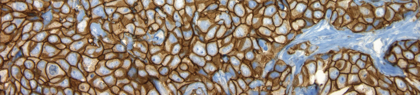
PATHWAY anti-HER-2/neu (4B5) Rabbit Monoclonal Primary Antibody (PATHWAY anti-HER2 (4B5) antibody) is a rabbit monoclonal antibody intended for laboratory use for the semi-quantitative detection of HER2 antigen by immunohistochemistry (IHC) in sections of formalin-fixed, paraffin-embedded normal and neoplastic breast tissue using the ultraView Universal DAB Detection Kit on a BenchMark ULTRA instrument.
This IHC device is indicated for identifying breast cancer patients who are eligible for treatment with Herceptin® (IHC 3+ or IHC 2+/ISH amplified), KADCYLA® (IHC 3+ or IHC 2+/ISH amplified) or ENHERTU® (IHC 1+ or IHC 2+/ISH non-amplified).
This product should be interpreted by a qualified pathologist in conjunction with histological examination, relevant clinical information, and proper controls.
This antibody is intended for in vitro diagnostic (IVD) use.
| Breast Cancer | Staining Pattern | Score | Recommended Reporting Status | Clinical Application |
|---|---|---|---|---|
No membrane staining is observed OR Faint, partial staining of the membrane in 10% or LESS of the cancer cells* |
0 | HER2 Negative | None | |
| Faint, partial staining of the membrane in greater than 10% of the cancer cells* | 1+ | HER2-low expression | ENHERTU® (fam-trastuzumab deruxtecan-nxki) |
|
| Weak to moderate complete staining of the membrane in greater than 10% of the cancer cells Positve Weak to moderate complete staining of the membrane in greater than 10% of the cancer cells Positve | 2+* Reflex test: HER2 Non-Amplified |
HER2-low expression | ||
2+* Reflex test: HER2 Amplified |
HER2 Positive/overexpression | Herceptin® (trastuzumab), KADCYLA® (trastuzumab emtansine) |
||
| Intense complete staining of the membrane in greater than 10% of the cancer cells | 3+ | HER2 Positive/overexpression |
Search our product catalogs and documentation library in eLabDoc.