Notre mission est d’améliorer la vie de tous les patients atteints de cancer. Pour ce faire et dans un souci d’innovation constante, nous développons pour le laboratoire de pathologie des instruments, des tests cliniques de cytologie, des tests cliniques sur tissus et des solutions logicielles, qui permettent aux anatomo-pathologistes du monde entier d’être plus performants.
Roche Tissue Diagnostics est le leader mondial des solutions qui aident les laboratoires de pathologie clinique à prévenir et diagnostiquer le cancer. Nous façonnons l’avenir des soins de santé personnalisés grâce à des instruments de pathologie intégrés, des tests cliniquement significatifs et des outils qui font progresser la certitude du diagnostic et optimisent la valeur médicale. Avec nos collègues de Roche et nos partenaires pharmaceutiques, nous développons et commercialisons également des tests prédictifs, qui permettent aux pathologistes et aux oncologues de cibler des traitements personnalisés pour chaque patient.
Des outils de diagnostic innovants pour la pathologie clinique
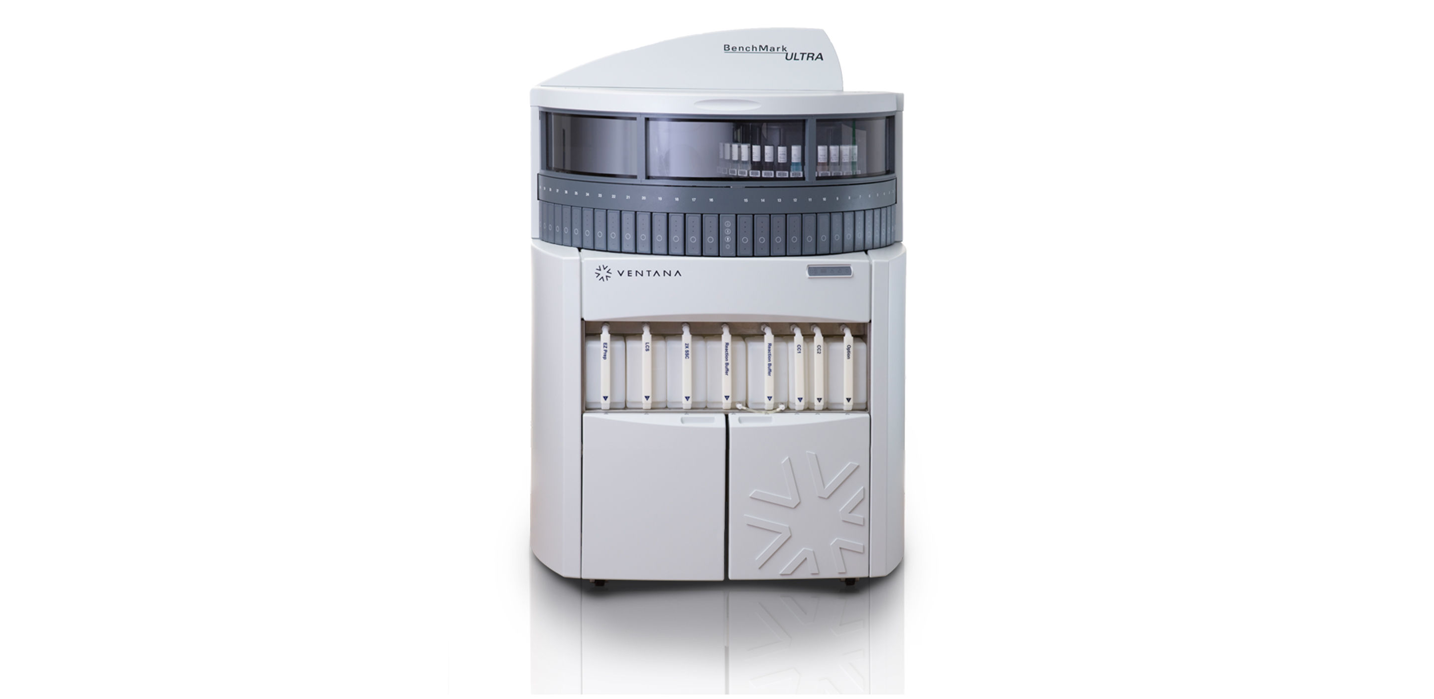
Notre portefeuille de produits VENTANA et BenchMark offre des résultats de cytologie et des résultats sur tissus rapides et précis pour que les pathologistes, les médecins et les patients puissent prendre des décisions de gestion clinique et de traitement en toute confiance. Notre équipement d’histologie comprend des plateformes automatisées de coloration H&E, d’immunohistochimie (IHC) et d’hybridation in situ (ISH) de nouvelle génération, ainsi que des kits de détection, des sondes et des tests cliniques extrêmement sensibles.
La solution de pathologie numérique de Roche associe du matériel (hardware) et des logiciels innovants, y compris la suite d’algorithmes pour analyse d’images uPath, ce qui permet de travailler de concert avec l’ensemble du portefeuille des diagnostics Roche et d’obtenir une solution complète. Roche fournit des solutions puissantes de pathologie numérique qui permettent des soins de santé améliorés et plus personnalisés, dès aujourd’hui et dans les années à venir.