Article

Evolving H&E - putting a trusted technique to the test

The hard-to-standardize standard in anatomical pathology

Making up about 80% of an anatomical pathology lab’s work, the hematoxylin and eosin (H&E) stain has become a mainstay as a rapid diagnostic for pathologists.


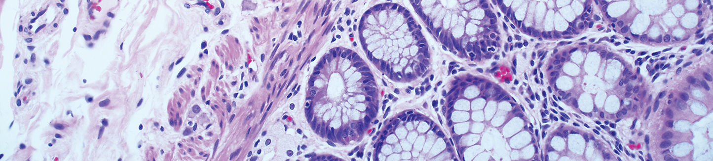
H&E is the principle laboratory method used to examine human tissue specimens for routine diagnosis, setting the course for all further work on patient tissue samples and directing the clinical course for patients. This technique of staining basophilic and acidophilic structures using hematoxylin and eosin, respectively, provides pathologists with functional and morphological details at the cellular level.1


Compared with other histological methods such as immunohistochemistry, H&E is simpler, faster, less expensive and more stable.1,2,3 However, it can be hard to standardize. The appearance and quality of each stain is dependent on numerous factors, such as the type of hematoxylin, the exact staining protocol, the quality of dyes, and their age.4 If the user is able to see what they need for interpretation, the quality is usually considered “satisfactory”, though this may vary by individual.


Despite the well-established utility of the technique, it has remained largely unchanged since its introduction more than a century ago, resulting in high variability between stains.

Striving for consistency—lab to lab and stain to stain

As understandings of oncology evolve, so too should testing.
 

The current manual, linear batch staining technique of H&E (also known as “dip and dunk”) has both procedural challenges and safety issues that may prevent accurate and timely patient diagnosis.
 

  • Patient cross-contamination may occur as tissue can dislodge from one slide and adhere to another5
  • Constant preparation of reagents and maintenance of solutions can lead to lack of consistent day-to-day staining and loss of time for histotechnicians that could be devoted to other functions2
  • Safety concerns around the use of hazardous chemicals such as xylene and alcohol6
     

People have preferences. Even within the same institution, differences in staining techniques may affect the quality or interpretation of a sample. Additionally, not all pathologists have the same training on the nuances of H&E staining.

The prevalence of protocol inconsistency and heavy reliance on “dip and dunk” processes present an opportunity to transform and modernize the way laboratories approach H&E staining.

Strengthening the “backbone” of pathology testing

Automation empowers laboratories to improve safety and quality with minimized process variability and standardized stains for pathologist review.
 

Consistent and confident interpretation enables a patient’s clinical course to be directed toward the best possible outcome and enables results to be more easily compared across laboratories and institutions.
 

Today, Roche offers the only automated H&E stainer that allows individual slide staining to empower high levels of workflow efficiency, stain quality, and safety. For patients, improved time to results, safe processing of their sample, and reliable identification and tracking help to ensure diagnosis continues as intended. For pathology labs, reducing hands-on time has the potential to:
 

  • Focus the efforts of laboratory personnel on value-added tasks
  • Reduce errors associated with manual handling
  • Virtually eliminate the potential for cross-contamination in H&E staining
  • Minimize operator contact with hazardous chemicals
     

As demand for testing grows, and as laboratory resources become increasingly scarce, standardized H&E staining with automation opens new possibilities for laboratory staff, pathologists and patients alike.

CTA background – microscopic tissue image collage

Special Stains—through increased efficiency

Specific staining provides information to better inform treatment decisions.

References

  1. Chan, J. The Wonderful Colors of the Hematoxylin–Eosin Stain in Diagnostic Surgical Pathology. Int J Surg Pathol. 2014;22(1):12–32.
  2. American Society for Mohs Histotechnology. Troubleshooting H&E staining. http://www.mohstech.org/UserFiles/file/Chow-TroubleshootHEStainingforMohs2017.pdf. Accessed 13 October 2020.
  3. Guo, L. Routine Hematoxylin and Eosin Stain Is Specific for the Diagnosis of Cytomegalovirus Infection in Gastrointestinal Biopsy Specimens. Int J Surg Pathol. 2018;26(6):500-506.
  4. Kleczek P, Jaworek-Korjakowska J, Gorgon M. A novel method for tissue segmentation in high-resolution H&E-stained histopathological whole-slide images. Comput Med Imaging Graph. 2020 Jan;79:101686.
  5. Cahill A, Pearson J. Measurement of stainer bath contamination and evaluation of common mitigation strategies. J Histotechnol. 2012;35(3):130-139.
  6. Feldman AT, Wolfe D. Tissue processing and hematoxylin and eosin staining. Methods Mol Biol. 2014;1180:31-43.