For localized information and support, would you like to switch to your country-specific website for {0}?
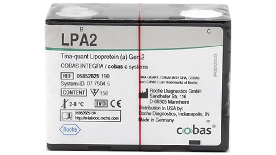
LPA2
Tina-quant Lipoprotein (a) Gen.2
IVD
For in vitro diagnostic use.
Use left and right arrow keys to scroll between the tabs
Overview
Technical documents
Access important product documentation including relevant certificates and other resources.
After clicking below, you will be redirected to eLabDoc, where you can select your local country-specific documents.

