For localized information and support, would you like to switch to your country-specific website for {0}?


Sustainability in diagnostics
Explore how Roche sustainability efforts are advancing sustainable healthcare through environmental, social and economic innovation across the diagnostics ecosystem.
Explore

ACT Ecolabels: Solving for transparency in sustainability
ACT ecolabels are now available for cobas® molecular reagents & assays for use on cobas® 5800/6800/8800 systems and the cobas® liat system and assay portfolio.
Explore
Featured products
Roche blood screening solutions deliver confidence in transfusion safety from donor to patient
IVD
For in vitro diagnostic use.
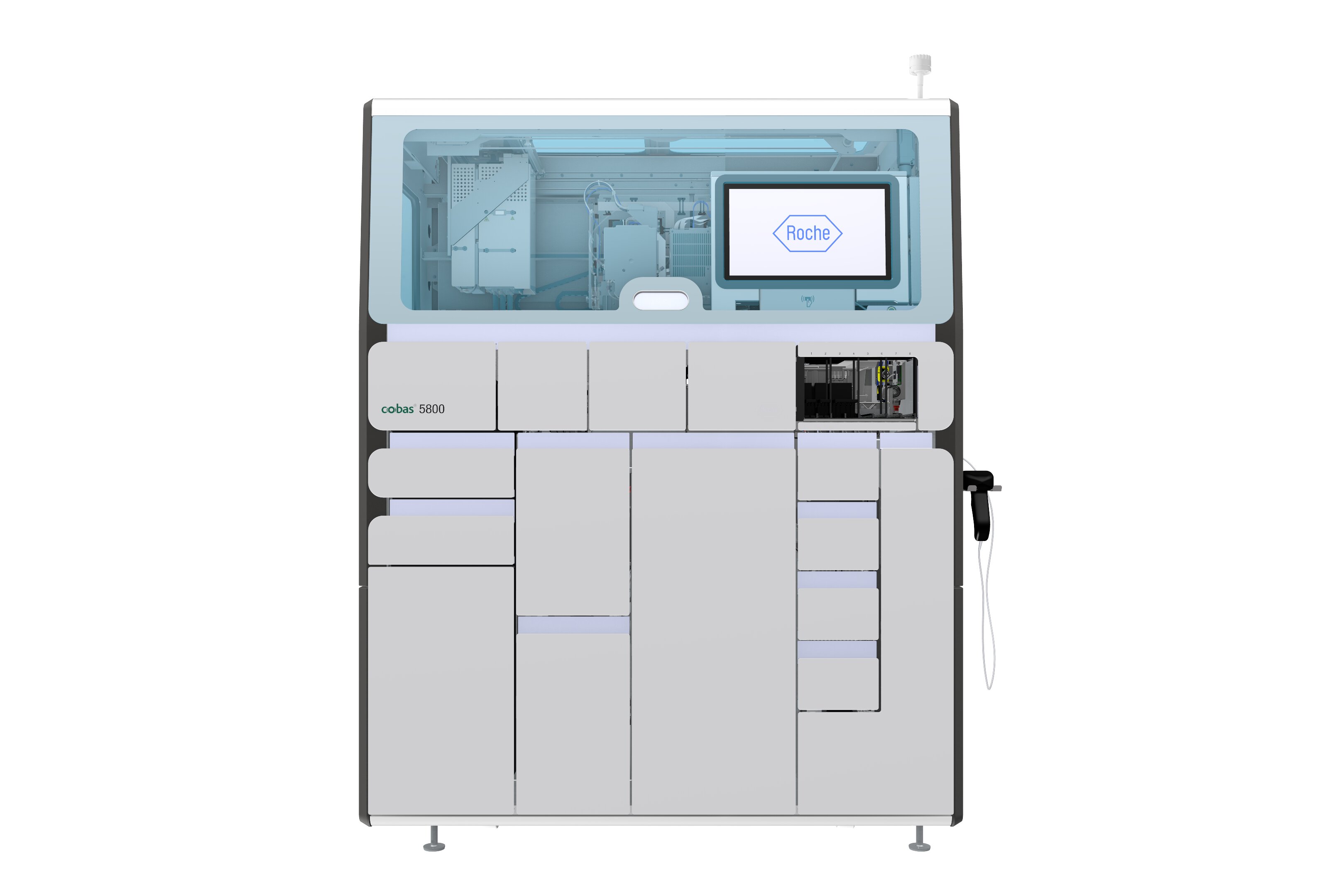
cobas® 5800 system
The cobas 5800 system supports an automated and integrated workflow to run PCR-based nucleic acid testing for use by trained professionals in laboratory settings.
IVD
For in vitro diagnostic use.
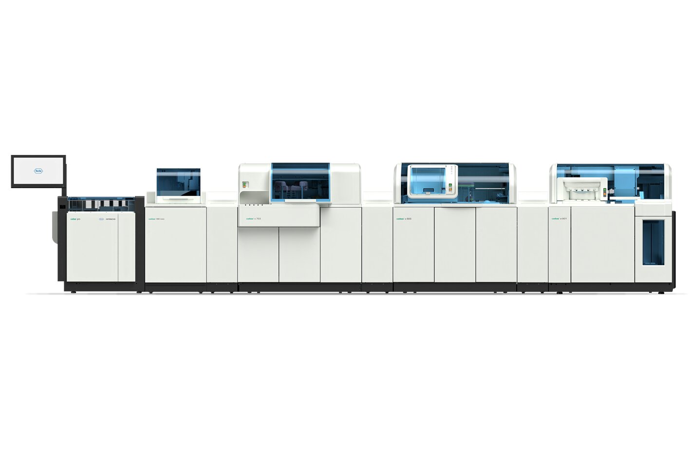
cobas® pro integrated solutions
The cobas® pro integrated solutions is a scalable and modular solution to achieve your mid to high-volume clinical chemistry and immunochemistry testing needs.
IVD
For in vitro diagnostic use.

cobas® pure integrated solutions
cobas pure integrated solutions combines clinical chemistry, immunoassay, and ion-selective electrode (ISE) testing on a footprint of just 2 square meters.
Integrated and customizable solutions powered by cutting-edge automation and digital technology.
IVD
For in vitro diagnostic use.
cobas® pro integrated solutions
The cobas® pro integrated solutions is a scalable and modular solution to achieve your mid to high-volume clinical chemistry and immunochemistry testing needs.
IVD
For in vitro diagnostic use.
cobas® pure integrated solutions
cobas pure integrated solutions combines clinical chemistry, immunoassay, and ion-selective electrode (ISE) testing on a footprint of just 2 square meters.
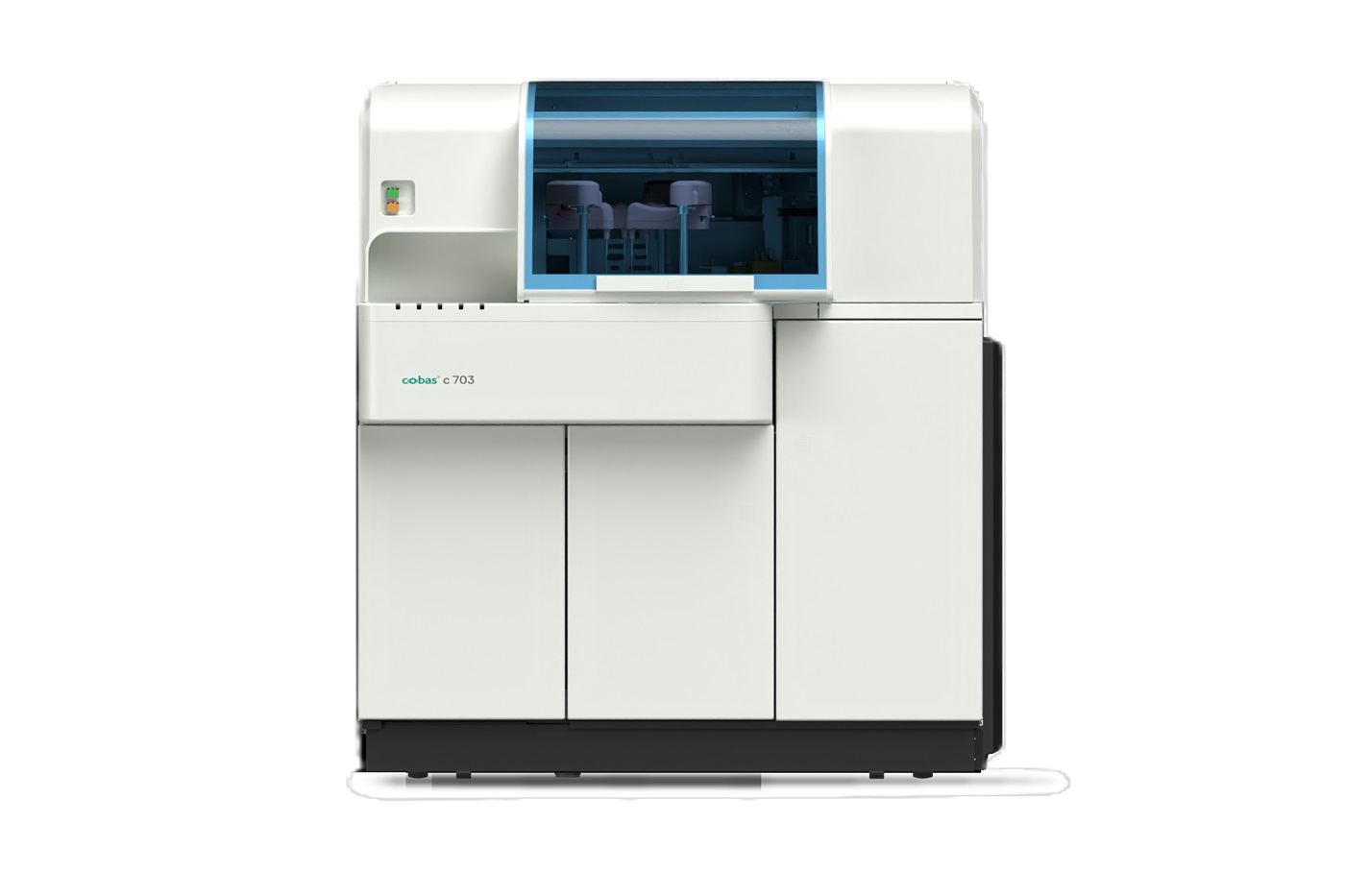
cobas® c 703 analytical unit
The cobas® c 703 analytical unit delivers high-throughput clinical chemistry testing, performing up to 2000 tests per hour and featuring 70 reagent positions.
A comprehensive portfolio of digital solutions designed to enhance operational, financial and clinical value, supporting informed decision-making in healthcare.
A comprehensive portfolio of molecular laboratory solutions—delivering confidence from samples in, to results out, across the continuum of healthcare.
IVD
For in vitro diagnostic use.
cobas® 5800 system
The cobas 5800 system supports an automated and integrated workflow to run PCR-based nucleic acid testing for use by trained professionals in laboratory settings.
IVD
For in vitro diagnostic use.
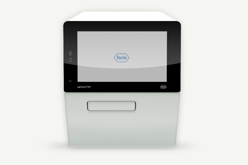
LightCycler® PRO system
LightCycler® PRO integrates innovative technology and Roche manufacturing to deliver accurate real-time PCR results
IVD
For in vitro diagnostic use.
cobas® eplex system
The cobas® eplex system delivers rapid, multiplex diagnostics with sample-to-answer automation, LIS integration, and scalable design.
Driving diagnostic certainty for life-changing decisions in pathology.
BenchMark ULTRA PLUS
The BenchMark ULTRA PLUS system’s fully-automated workflow for slide staining improves turnaround time and decreases touchpoints.
Country Specific Labeling
See country-specific product labeling for regulatory status.
VENTANA® DP 600 slide scanner
The VENTANA DP 600 slide scanner enables full digitization of your pathology lab by offering high-volume and high-quality whole slide scanning.
IVD
For in vitro diagnostic use.
BenchMark Special Stains
The BenchMark Special Stains system automatically stains histology specimens with reagents for in vitro diagnostics, enhancing workflow with proven technology.
Solutions that empower you to provide personalized medicine and shape a future of preventive, coordinated, and value-based care—everywhere it’s needed.
IVD
For in vitro diagnostic use.
cobas® h 232 POC system
The cobas h 232 is an in vitro diagnostic instrument intended for the quantitative evaluation of immunoassays using the Roche CARDIAC test strips in human whole blood. The cobas h 232 is intended for near-patient testing. Not for self-testing.
IVD
For in vitro diagnostic use.
cobas® liat system
Explore the cobas® liat system from Roche, gold-standard PCR technology at the Point of Care and see how you can elevate patient care.
IVD
For in vitro diagnostic use.
cobas® pulse system
Explore the cobas® pulse professional glucose system from Roche Diagnostics, providing advanced performance and a wide range of selected mobile health apps.
Efficient, high-performance solutions for next-generation sequencing (NGS).
RUO
For Research Use Only. Not for use in diagnostic procedures.
AXELIOS 1 Platform
Discover AXELIOS 1. A next generation sequencing platform powered by SBX technology. Deliver faster results, flexible throughput, and scalable workflows for genomics research and clinical research applications.
RUO
For Research Use Only. Not for use in diagnostic procedures.
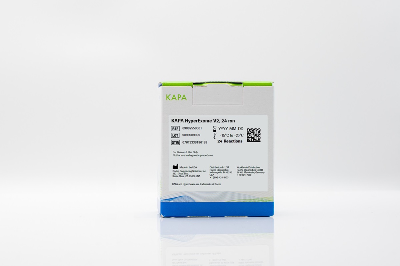
KAPA HyperExome V2 Probes and Kits
The KAPA HyperExome V2 Probes are Roche's brand new Whole Exome Sequencing solution delivering superior content coverage.
RUO
For Research Use Only. Not for use in diagnostic procedures.
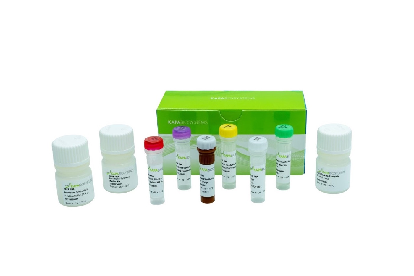
KAPA RNA EvoPrep Kits
Maximize Every Read. Our new KAPA RNA EvoPrep Kit shortens workflow, boosts sequencing efficiency, captures more unique transcripts, and accelerates discovery.

Insights


?wid=384&hei=216&fmt=png-alpha&fit=crop,1&cropn=0.07,0,0.86,1)


