You appear to be using incognito/private browsing mode or an ad blocker, which may adversely affect your experience on the site. Please disable any ad blockers and view the site in non-private mode.
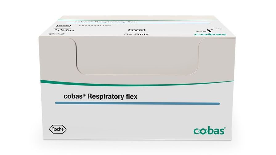
For in vitro diagnostic use. Others cobas Respiratory flex IVD cobas® Respiratory flex PID00000771 Qualitative nucleic acid test for use on the cobas® 5800/6800/8800 systems 09 623 701 190 9 623 701 190 09623701190 9623701190 09623701190 KIT COBAS 58/68/8800 RESP FLEX 192T IVD KIT COBAS 58/68/8800 RESP FLEX 192T IVD 00875197007312 Reagents, kits 1 kit 192 tests true cobas® Respiratory flex for use on the cobas® 5800/6800/8800 systems (cobas® Respiratory flex) is an automated, multiplex, nucleic acid test that utilizes real-time polymerase chain reaction (PCR) technology for simultaneous in vitro qualitative detection and differentiation of adenovirus (species B, C and E), common human coronaviruses (229E, HKU1, NL63, OC43), human metapneumovirus, human rhinovirus/enterovirus, influenza A virus, influenza B virus, parainfluenza viruses 1, 2, 3, and 4, respiratory syncytial virus (RSV), and SARS-CoV-2 in nasopharyngeal swab specimens obtained from individuals with signs and symptoms of respiratory tract infections in conjunction with clinical and epidemiological risk factors.
The detection and identification of specific viral nucleic acids from individuals presenting with signs and symptoms of a respiratory infection aids in the diagnosis of respiratory infection if used in conjunction with other clinical and epidemiological information. Negative results do not preclude a respiratory infection and should not be used as the sole basis for treatment or other patient management decisions. Conversely, positive results do not rule out coinfection with other organisms, and the agent detected may not be the definite cause of disease.
Due to the genetic similarity between human rhinovirus and enterovirus, the cobas® Respiratory flex test cannot reliably differentiate them. en cobas® Respiratory flex is based on fully automated sample preparation (nucleic acid extraction and purification) followed by PCR amplification and detection. The cobas® 5800 System is designed as one integrated instrument. The cobas® 6800/8800 systems consist of the sample supply module, the transfer module, the processing module, and the analytic module. Automated data management is performed by the cobas® 5800 system or cobas® 6800/8800 systems software(s), which assigns results for all tests.
Results can be reviewed directly on the system screen, and printed as a report.
Nucleic acid from patient samples and added Internal Control RNA (RNA IC) molecules are simultaneously extracted. Nucleic acid is released by addition of proteinase and lysis reagent to the sample. The released nucleic acid binds to the silica surface of the added magnetic glass particles. Unbound substances and impurities, such as denatured protein, cellular debris and potential PCR inhibitors, are removed with subsequent wash steps and purified nucleic acid is eluted from the magnetic glass particles with elution buffer at elevated temperature. External controls (positive and negative) are processed in the same way.
Selective amplification of target nucleic acid from the sample is achieved by the use of target-specific forward and reverse primers detecting conserved viral genome regions.
Targeted
Target gene (symbol)
Influenza A
Matrix protein 1 (M1)
Influenza B
Non-structural protein NS-1/2 (NS1/NEP)
Respiratory Syncytial Virus
Matrix protein (M)
SARS-CoV-2
ORF1 ab polyprotein (ORF1ab) and ORF 1a polyprotein (ORF1a)
Adenovirus B/E
Terminal protein precursor (E2B)
Adenovirus C
Capsid protein precursor (L3)
Human Metapneumovirus
Matrix protein (M)
Enterovirus/Rhinovirus
5’ untranslated region (5’UTR)
Coronavirus OC43
Replicase polyprotein 1 a/b (ORF1ab
Coronavirus HKU1
Replicase polyprotein 1 a/b (ORF1ab)
Coronavirus 229E
Replicase polyprotein 1 a/b (ORF1ab)
Coronavirus NL63
Replicase polyprotein 1 a/b (ORF1ab)
Human Parainfluenza Virus 1
L polymerase protein (L)
Human Parainfluenza Virus 2
Large protein (L)
Human Parainfluenza Virus 3
Nucleocapsid protein (N)
Human Parainfluenza Virus 4
Large protein (L)
Selective amplification of RNA IC is achieved by the use of non-competitive, sequence specific forward and reverse primers, which have no homology with the viral-target specific genomes. Amplified target is detected by the cleavage of fluorescently labeled oligonucleotide probes. Roche’s temperature assisted generation of signal (TAGS) technology, short TAGS technology, is introduced to differentiate up to three targets per fluorescence channel, enabling the detection of 12 targets, and the Internal Control, per well. A thermostable DNA polymerase enzyme is used for amplification.
The cobas® Respiratory flex master mix contains detection probes which are specific for adenovirus (species B,C and E), common human coronaviruses (229E, HKU1, NL63, OC43), human metapneumovirus, human rhinovirus/enterovirus, influenza A virus, influenza B virus, parainfluenza viruses 1, 2, 3, and 4, RSV, and SARS-CoV-2, and the RNA Internal Control nucleic acid. Multiplicity of target detection is enabled with temperature-dependent quenching of cleaved fluorescent target-specific probes. This is achieved by separating signals from probes into introduced thermal channels, where fluorescence is acquired at two additional fixed temperatures for each amplification cycle.
During the PCR amplification step, hybridization of the probes to the specific single-stranded DNA template results in cleavage of the probe by the 5' to 3' exonuclease activity of the DNA polymerase, resulting in separation of the reporter and quencher dyes, and the generation of a fluorescent signal. Conventional probes release fluorescence signal immediately upon separation of reporter from quencher. TAGS probes rely on temperature dependent fluorescence activation, requiring both nuclease cleavage during the extension phase, as well as an increase in reaction temperature, to activate the otherwise dormant fluorophore. For this reason, during each PCR cycle the test captures fluorescence in five available fluorescence channels in combination with three thermal channels (detection of fluorescence at three defined temperatures T1 T2 and T3), which enables simultaneous detection and differentiation of the amplified adenovirus (species B,C and E), common human coronaviruses (229E, HKU1, NL63, OC43), human metapneumovirus, human rhinovirus/enterovirus, influenza A virus, influenza B virus, parainfluenza viruses 1, 2, 3, and 4, RSV, and SARS-CoV-2 viral targets and the RNA IC.
The master mix includes deoxyuridine triphosphate (dUTP), instead of deoxythymidine triphosphate (dTTP), which is incorporated into the newly synthesized DNA (amplicon). Any contaminating amplicons from previous PCR runs are destroyed by the AmpErase enzyme [uracil-N-glycosylase], which is included in the PCR mix, when heated in the first thermal cycling step. However, newly formed amplicons are not destroyed since the AmpErase enzyme is inactivated once exposed to temperatures above 55°C.
en