Breast cancer IHC/ISH portfolio


Deliver diagnostic confidence with the Roche breast cancer IHC/ISH portfolio
As a leader in breast cancer diagnostics, Roche Diagnostics offers numerous cancer diagnostic assays that enable pathology labs to deliver accurate results with confidence. We provide a robust menu of breast cancer diagnostic tools. Our portfolio of products delivers the high sensitivity and specificity you need from your assays.
Immunohistochemistry (IHC) and in situ hybridization (ISH)
Experts predict over 680,000 deaths annually due to breast cancer, making it a leading cause of death in women.1 In developed countries breast cancer-related mortality has declined, in part due to therapeutic advances and evolution of biomarkers.2,3 For example, Immunohistochemistry (IHC) analysis and in situ hybridization (ISH) analysis provide important information related to tumor biology, help profile types of breast cancer and may improve treatment decisions.4
Clinical benefit
With proven accuracy, VENTANA breast cancer diagnostic assays help you identify patients other assays can miss—so you can deliver the right test, with clinical confidence in the shortest time possible.
Analytical advantage
Specific and sensitive rabbit monoclonal antibodies, best-in-class probes and powerful detection systems help you diagnose precisely and confidently.
Testing efficiency
Our comprehensive breast cancer workflow solution delivers fully automated assays on market-leading platforms, with digital pathology and workflow solutions that free up resources, reduce labor costs and reduce time to results.
Featured assays
CONFIRM ER (SP1) Rabbit Monoclonal Primary Antibody
- FDA cleared and CE IVD antibody
- Indicated as an aid in patient management, prognosis, and the prediction of therapy outcomes in breast cancer.5
- ER (SP1) is a significant predictor of disease-specific survival.6,7
- Rapid and consistent results delivered through fully automated platforms and digital pathology solutions

CONFIRM PR (1E2) Rabbit Monoclonal Primary Antibody
- FDA cleared and CE IVD antibody
- Indicated as an aid in patient management, prognosis, and the prediction of therapy outcomes in breast cancer.14
- Provides significant value as a prognostic factor and response prediction of hormone therapy, even in ER negative patients.10
- Rapid and consistent results through fully automated platforms15

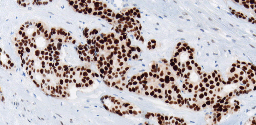
CONFIRM anti-Ki-67 (30-9) Rabbit Monoclonal Primary Antibody
- Aids in assessing the proliferative activity of normal and neoplastic cells16,17,18
- Rabbit monoclonal antibody demonstrates increased sensitivity and strong specificity compared with mouse monoclonal antibodies19
- Intense nuclear staining and no adipose (K2) or cell membrane staining can help deliver a confident assessment of tumor aggressiveness20

PATHWAY HER2 (4B5) Rabbit Monoclonal Primary Antibody
- FDA approved and CE IVD antibody
- Higher overall proficiency assessment scores with HER2 (4B5) than with any other clones11
- Widely adopted and reliable HER2 IHC primary antibody6
- High concordance with HER2 ISH12,13

VENTANA HER2 Dual ISH DNA Probe Cocktail assay
- FDA approved and CE IVD test
- Increased performance with oligo probes and new detection kits
- Easily interpreted using brightfield microscopy
- Robust and highly reproducible between laboratories and pathologists8
- Highly concordant with FISH8

VENTANA PD-L1 (SP142) Assay
- Reliably detect PD-L1 expression on immune cells21
- Confidently identify TNBC patients eligible for treatment22
- Enable care teams to chart new courses for personalized breast cancer treatment

References
- WHO Globocan 2020 Breast Cancer Fact Sheet. https://gco.iarc.fr/today/data/factsheets/cancers/20-Breast-fact-sheet.pdf
- Berry DA, Cronin KA, Plevritis SK, et al. Effect of screening and adjuvant therapy on mortality from breast cancer. N Engl J Med. 2005;353:1784‐1792.
- Munoz D, Near AM, van Ravesteyn NT, et al. Effects of screening and systemic adjuvant therapy on ER‐specific US breast cancer mortality. J Natl Cancer Inst. 2014;106:dju289.
- Godoy-Ortiz, A. et al. Deciphering HER2 Breast Cancer Disease: Biological and Clinical Implications. Front. Oncol. 2019;9:1124
- CONFIRM anti-Estrogen Receptor (ER) (SP1) Rabbit Monoclonal Primary Antibody package insert, 2013.
- Welsh, A. et al. Quantitative Analysis of Estrogen Receptor Expression Shows SP1 Antibody Is More Sensitive Than 1D5. Appl Immunohistochem Mol Morphol.2013; 21(2):139-147.
- Yamamoto-Ibusuki M. et al. Comparison of prognostic values between combined immunohistochemical score of estrogen receptor, progesterone receptor, human epidermal growth factor receptor Ki-67 and the corresponding gene expression score in breast cancer. Mod Patholo 2013:26:79-86.
- VENTANA HER2 Dual ISH DNA Probe Cocktail Assay package insert, 2020.
- Lim, SJ. et al. Validation and workflow optimization of human epidermal growth factor receptor 2 testing using INFORM HER2 dual-color in situ hybridization. Human Pathology. 2013; 44, 2590–2596.
- Ruschoff, J. et al. ISH-based HER2 diagnostics. Pathologe. 2020;(6):606-613.
- Based on 5 years of data from a leading external quality assessment scheme. B30 2020 HER2 IHC assessment run. Retrieved from http/www.nordiqc.org/epitopes.htm.
- Mayr D. et al. Comprehensive immunohistochemical analysis of Her-2/neu oncoprotein overexpression in breast cancer: HercepTest (Dako) for manual testing and Her-2/neu Test 4B5 (VENTANA) for VENTANA BenchMark automatic staining system with correlation to results of fluorescence in situ hybridization (FISH). Virchows Archiv.2009:454(3):241-248.
- Brugmann A. Lelkaitis G. Nielsen S. et al. Testing HER2 in breast cancer: a comparative study on BRISH, FISH and IHC. Appl Immunohistochem Mol Morphol. 2011:19(3):203-211.
- CONFIRM anti-Progesterone Receptor (PR) (1E2) Rabbit Monoclonal Primary Antibody package insert, 2012.
- Liu, S. Chia SK. Mehl, E. et al. Progesterone receptor is a significant factor associated with clinical outcomes and effect of adjuvant tamoxifen therapy in breast cancer patients. Breast Cancer Res Treat 2010:119:53-61.
- Keng PC, Siemann DW. Measurement of proliferation activities in human tumor models: a comparison of flow cytometric methods. Radiatn Oncol Investig. 1998: 6(3): 120-127.
- Rey A Lara PC. Ki67 proliferation index in tumors of the upper urinary tract as related to established prognostic factors and long-term survival. Arch Esp Urol. 1998.51(2): 204-210.
- Bacchi CE, Gown AM. Detection of cell proliferation in tissue sections. 1993.Braz J Med Biol Res, 26(7): 677-87.
- Minca, E. et al. Automated and Visual Assessment of Ki-67 IHC in Breast Carcinoma: Comparison of 3 Antibody Clones. Laboratory Investigation, suppl. 2012; 93:57A pg. 33.
- Leonardo E, Volante M, Barbareschi M, Cavazza A, Dei TAP, Bussolati G, et al. Cell membrane reactivity of MIB-1 antibody to Ki67 in human tumors: fact or artifact? Appl Immunohistochem Mol Morphol.2007; 15(2): 220–223.
- VENTANA PD-L1 (SP142) Assay Package Insert, 2019.
- Schmid P, Adams S, Rugo HS, et al. Atezolizumab and Nab-Paclitaxel in Advanced Triple-Negative Breast Cancer. N Engl J Med. 2018;379(22):2108-2121.