VENTANA PD-L1 (SP263)

VENTANA PD-L1 (SP263) Assay, non-small cell lung cancer (NSCLC), urothelial carcinoma, bladder cancer
The VENTANA PD-L1 (SP263) assay is now FDA approved to detect PD-L1 across NSCLC to identify patients eligible for multiple targeted therapies.

The VENTANA PD-L1 (SP263) Assay is an FDA-approved CDx that enables your lab to identify NSCLC patients eligible for treatment with TECENTRIQ (atezolizumab)  in early stage NSCLC.

The VENTANA PD-L1 (SP263) Assay is an FDA-approved CDx that enables your lab to identify NSCLC patients eligible for treatment with LIBTAYO (cemiplimab-rwlc) in locally advanced or metastatic NSCLC.

Ordering information

VENTANA PD-L1 (SP263) Assay
Material Number: 07208162001
Part Number: 740-4907

Additional education

VENTANA PD-L1 (SP263) Assay Slide Review
VENTANA PD-L1 (SP263) Assay Overview
VENTANA PD-L1 (SP263) In NSCLC
VENTANA PD-L1 (SP263) 25% PD-L1 Expression
VENTANA PD-L1 (SP263) educational video


VENTANA PD-L1 (SP263) Rabbit Monoclonal Primary Antibody Class I

PD-L1 is an informative immunotherapy biomarker: Aberrant expression of PD-L1 on tumor and immune cells

in the tumor microenvironment impedes anti-tumor immunity, allowing tumors to grow and metastasize.

Broad Tissue Application

Aids in PD-L1 Expression for Many Tumor Types

The VENTANA PD-L1 (SP263) Rabbit Monoclonal Primary Antibody is intended for laboratory use in the PD-L1 protein in the formalin-fixed, paraffin-embedded tissue. It is intended to be used on the BenchMark Ultra. It is indicated as an aid in the assessment of PD-L1 expression in human tissues.

  • Interruption of the PD-L1/PD-1 pathway represents an attractive strategy to reinvigorate tumor-specific T cell immunity suppressed by the expression of PD-L1 in the tumor microenvironment.
  • PD-L1 is expressed in a broad range of cancers including lung, melanoma, urothelial, ovarian, head and neck, renal, hepatocellular, gastric or gastroesophageal junction, and colorectal cancer.
fda-approved-image

*In the US only available on the BenchMark ULTRA Instrument

PD-L1 (SP263)
Learn More about the VENTANA PD-L1 (SP263) Rabbit Monoclonal Primary antibody assay Class I

VENTANA PD-L1 (SP263) Rabbit Monoclonal Antibody Class I Immune Cell Scoring Video Series

Part I
Part II
Part III

Class I
Class I Product Information
Package Insert

Ordering information

VENTANA PD-L1 (SP263) Rabbit  Monoclonal Antibody
Material Number: 07494190001
Part Number: 790-4905

Would you like to know more about VENTANA PD-L1 (SP263)?

Please submit your information in the following form to be contacted by a Roche representative with more details.

Form Successfully Submitted!
Thank you for your submission!
text

The PD-L1 immunologic checkpoint

About PD-L1

PD-L1 is a transmembrane protein that down-regulates immune responses through binding to its two inhibitory receptors, programmed death-1 (PD-1) and B7.1. PD-1 is an inhibitory receptor expressed on T cells following T-cell activation, which is sustained in states of chronic stimulation such as in chronic infection or cancer.1 Binding of PD-L1 with PD-1 inhibits T cell proliferation, cytokine production and cytolytic activity, leading to the functional inactivation or exhaustion of T cells. B7.1 is a molecule expressed on antigen presenting cells and activated T cells. PD-L1 binding to B7.1 on T cells and antigen presenting cells can mediate down-regulation of immune responses, including inhibition of T-cell activation and cytokine production.2 PD-L1 expression has been observed in immune cells and tumor cells.3,4 Aberrant expression of PD-L1 on tumor cells has been reported to impede anti-tumor immunity, resulting in immune evasion.1 Therefore, interruption of the PD-L1/PD-1 pathway represents an attractive strategy to reinvigorate tumor-specific T cell immunity suppressed by the expression of PD-L1 in the tumor microenvironment.

checkpoint-image

References

  1. Blank, C and Mackensen, A, Contribution of the PD-L1/PD-1 pathway to T-cell exhaustion: an update on implications for chronic infections and tumor evasion. Cancer Immunol Immunother, 2007. 56(5): p. 739-745. 
  2. Butte MJ, Keir ME, Phamduy TB, et al. Programmed death-1 ligand 1 interacts specifically with the B7-1 costimulatory molecule to inhibit T cell responses. Immunity. 2007;27(1):111-122. 
  3. Dong H, Zhu G, Tamada K, Chen L. B7-H1, a third member of the B7 family, co-stimulates T-cell proliferation and interleukin-10 secretion. Nat Med. 1999;5(12):1365-1369. 
  4. Herbst RS, Soria JC, Kowanetz M, et al. Predictive correlates of response to the anti-PD-L1 antibody MPDL3280A in cancer patients. Nature. 2014;515(7528):563-567.

The tumor microenvironment

Plays a key role in tumor progression
 

What is the tumor microenvironment (TME)?

The tumor microenvironment (TME) consists of different cellular, including immune cells, and non-cellular components in and around the tumor. The TME has been recognized to play a significant role in tumor progression.1,2
 

Why is the TME important?

The TME shapes tumor evolution (whether the tumor regresses, develops resistance, evades the immune system and/or metastasizes) and consequently impacts patient outcomes.3 An association has been observed between the levels of tumor infiltrating immune cells, key components of the TME, and patient prognosis: a colorectal cancer study showed that higher levels of tumor infiltrating CD3+ immune cells were associated with better disease free survival.4
 

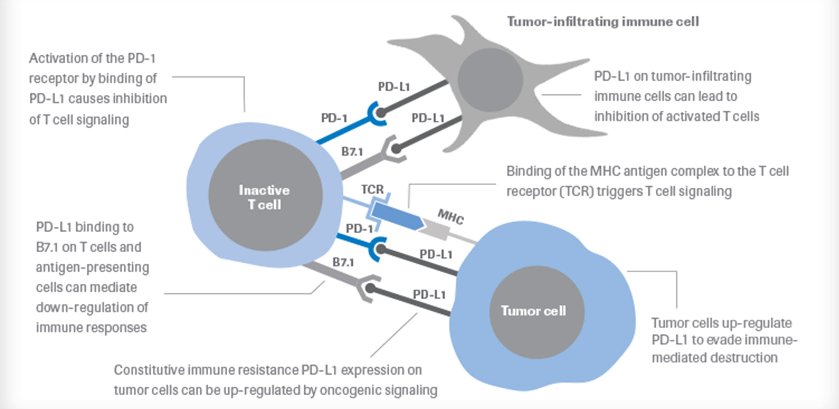
What is the role of PD-L1 in the TME?

Aberrant expression of PD-L1 on tumor cells has been reported to impede anti-tumor immunity, resulting in immune evasion.5 Therefore, interruption of the PD-L1/PD-1 pathway represents an attractive strategy to reinvigorate tumor-specific T cell immunity suppressed by the expression of PD-L1 in the TME. This approach has proven effective.

microenvironment-image

References 

  1. NCI Dictionary of Cancer Terms. Tumor microenvironment. http://www.cancer.gov/publications/dictionaries/cancer-terms?cdrid=561725 
  2. Hinshaw DC and Shevde LA. The tumor microenvironment innately modulates ccancer progression. Cancer Research 2019; 79: 4557-66. 
  3. Chen F, Zhuang X, Lin L, Yu P, Wang Y, Shi Y, Hu G, Sun Y.BMC Med. New horizons in tumor microenvironment biology: challenges and opportunities. 2015 Mar 5;13:45. doi: 10.1186/s12916-015-0278-7. 
  4. Galon J1, Costes A, Sanchez-Cabo F, Kirilovsky A, Mlecnik B, Lagorce-Pagès C, Tosolini M, Camus M, Berger A, Wind P, Zinzindohoué F, Bruneval P, Cugnenc PH, Trajanoski Z, Fridman WH, Pagès F. Type, density, and location of immune cells within human colorectal tumors predict clinical outcome. Science. 2006 Sep 29;313(5795):1960-4. 
  5. Blank, C and Mackensen, A, Contribution of the PD-L1/PD-1 pathway to T-cell exhaustion: an update on implications for chronic infections and tumor evasion. Cancer Immunol Immunother, 2007. 56(5): p. 739-745). 
  6. Ventana Medical Systems Inc. VENTANA PD-L1 (SP142) Assay. Package Insert. http://www.ventana.com/product/1827?type=2357

Research Use Only

uPath


Learn More about the RUO PD-L1 (SP263) Image Analysis Algorithm

RUO: SP263 Image Analysis Algorithm Video
Intro to uPath PD-L1 SP263 Image Analysis Algorithm


NEW: Roche uPath Image Analysis Algorithms
PD-L1 (SP263) Image Analysis NSCLC Brochure

SP263